.svg)
.svg)

.svg)

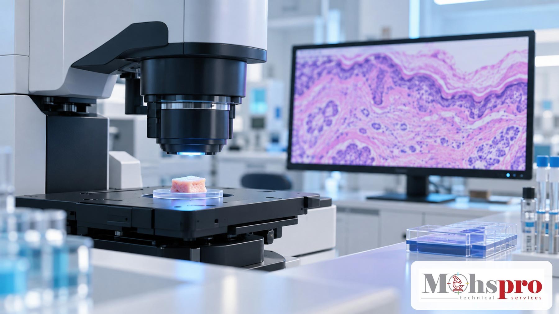
A new study reports encouraging accuracy for two photon fluorescence microscopy on fresh Mohs specimens while underscoring the need for prospective intraoperative validation.
Article content
A study published in the September 2026 issue of the Journal of the American Academy of Dermatology evaluated whether two photon fluorescence microscopy could provide H and E colored images of fresh Mohs tissue without conventional slide preparation. The findings are promising, but they represent an investigational workflow rather than a replacement for frozen section histology.
What the Researchers Evaluated
The investigators acquired conventional frozen sections and two photon fluorescence microscopy images from 133 specimens collected after completion of Mohs surgery for basal cell carcinoma. Two Mohs surgeons who had not previously interpreted this type of microscopy reviewed the digital images. Their readings were compared with a consensus frozen section assessment made by a dermatopathologist and a separate Mohs surgeon.
Two photon fluorescence microscopy is a slide free optical method that can generate H and E like images from fresh tissue. In principle, eliminating freezing, sectioning, staining, mounting, and slide handling could reduce several manual steps. That possibility makes the technology relevant to practices interested in faster intraoperative evaluation and new digital pathology workflows.
What the Study Found
Across the study specimens, the average sensitivity reported for the two readers was 93.8 percent and the average specificity was 82.2 percent. When the investigators excluded discordant specimens attributed to differences in the tissue plane or to fragments produced during frozen sectioning, the averages increased to 95.0 percent sensitivity and 94.3 percent specificity.
Those adjusted values require careful interpretation. The exclusions were based on discrepancies between what was present in the optical image and what appeared on the corresponding frozen section. They help describe the effect of sampling and sectioning differences, but they should not be treated as the primary real world performance estimate.
Why This Matters for Mohs Practices
Frozen section histology remains central to current Mohs workflows. Producing complete, interpretable sections depends on accurate orientation, controlled embedding, consistent section thickness, effective staining, and traceable specimen handling. A slide free system could change where time and quality risks occur, moving part of the workflow from cryotomy and staining toward tissue positioning, image acquisition, digital quality control, and system validation.
The study also suggests that Mohs surgeons may be able to learn the H and E colored optical appearance with limited prior exposure. However, the research did not test the technology as the sole basis for intraoperative decisions during active surgery. It therefore does not establish that a laboratory should remove conventional frozen section steps or alter validated procedures.
For histotechnologists, the more immediate lesson is that emerging imaging systems will still depend on excellent specimen orientation, complete surface presentation, documentation, and prompt escalation when tissue or image quality is inadequate. Technology may change the instruments, but it does not eliminate the need for disciplined quality control.
Study Limitations
The study focused on basal cell carcinoma specimens and evaluated agreement after Mohs surgery had concluded. It did not prospectively measure patient outcomes, stage completion time, workflow failures, cost, training requirements, or performance across a broader range of tumors and artifacts. The authors reported no conflicts of interest. The work received support from National Institutes of Health grants listed in the publication.
Key Takeaways
- Two photon fluorescence microscopy generated H and E colored images from fresh Mohs specimens and achieved encouraging diagnostic agreement in this study.
- The strongest adjusted results depended on excluding specimens affected by tissue plane differences or frozen section fragments.
- Prospective intraoperative validation is still needed before the technology can be considered an alternative to standard frozen section histology.
Tips for Histotechnologists
Mohs workflow
- Document specimen orientation before processing so the intended margin can be correlated with the image or slide.
- Confirm complete epidermal and deep margin representation and escalate folds, gaps, chatter, or missing tissue before interpretation.
- Maintain traceability between the map, specimen, block, slide, and any digital image generated during the case.
Histopathology workflow
- Verify stain quality and control performance before accepting a run for review.
- Record recuts, levels, repeated stains, and corrective actions in the laboratory record.
- Escalate image quality, tissue adequacy, or instrument performance concerns to the responsible pathologist or laboratory supervisor.
Educational Disclaimer
This article is for professional education and does not provide individual medical advice or recommend changes to validated laboratory or clinical procedures.

.svg)
.svg)