.svg)
.svg)

.svg)

A new observational study tested bioelectrical impedance and machine learning to map nodular basal cell carcinoma risk during Mohs surgery. The technology produced rapid probability maps, but frozen-section histology remained the definitive assessment and important validation questions remain.
Article content
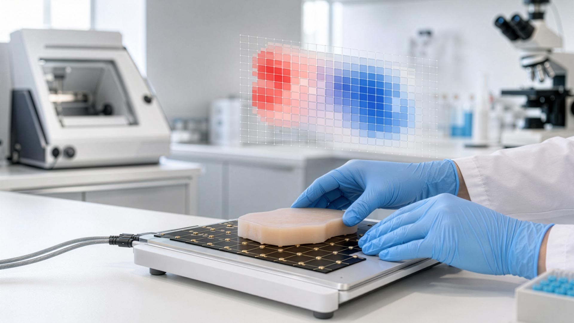
A study published August 11, 2026, in npj Biomedical Innovations evaluated whether high-frequency bioelectrical impedance spectroscopy combined with supervised machine learning could provide rapid, spatial guidance during Mohs surgery. The system generated probability heat maps from excised specimens before routine frozen-section processing. It was studied only as an adjunct: its output was not used for clinical decisions, and frozen-section histopathology remained the definitive method for determining margin clearance.
How the Experimental System Worked
Bioelectrical impedance spectroscopy measures how tissue responds to alternating electrical signals. Because electrical properties can reflect tissue composition and microstructure, the researchers evaluated whether a machine-learning model could distinguish locations associated with nodular basal cell carcinoma from locations labeled negative by histopathology.
The IRB-approved, ex vivo observational study included 98 valid specimens from 55 patients with nodular BCC. Eighteen specimens with non-nodular BCC patterns and two specimens for technical reasons were excluded from machine-learning development. After measurement on an electrode array, routine frozen sections were prepared and interpreted. Histopathology findings were then translated onto a digital grid to create reference labels for model training and evaluation.
What the Study Reported
Under strict location-by-location scoring, the cancer classifier achieved a mean receiver operating characteristic area under the curve (ROC AUC) of 0.878 ± 0.048. At the reported F1 operating threshold, the negative predictive value was 94.9%, but 90 positive locations were classified as false negatives. The paper also reported a separate spatial-tolerance analysis intended to account for deformation and registration error between the fresh specimen, the electrode grid, and the later frozen-section map.
With that tolerance-based evaluation, the reported mean ROC AUC was 0.993 ± 0.003, with sensitivity of 95.6% ± 4.7% and specificity of 96.1% ± 5.0%. Those higher values must be described together with the scoring method: the spatial tolerance was an evaluation adjustment for observed map misregistration, not a prospectively validated universal clinical tolerance.
Median acquisition time was estimated at 3.7 minutes per specimen (interquartile range, 2.6–5.5 minutes), plus an estimated two minutes for gel and array preparation. The authors described the measurement as non-destructive, allowing the specimen to continue through the standard frozen-section workflow.
Why This Matters for Mohs Practices
The potential value is timing. A rapid probability map could, in principle, give the surgeon early regional guidance while histology is still being processed. That might support planning or workflow coordination in busy practices. The study does not show that the device reduces stages, patient waiting time, overtime, errors, or costs in real-world clinical use; those outcomes require prospective evaluation.
For histotechnologists, the study also highlights a central challenge: exact correlation between a fresh specimen and its final microscopic representation is difficult. Positioning on an array, lifting, embedding, freezing, sectioning, slide transfer, annotation, and digitization can each alter geometry. Reliable orientation, complete sections, clear mapping, and traceable documentation therefore remain essential even when adjunctive mapping technology is added.
Important Limitations and Conflicts
This was a small, single-center observational study limited to nodular BCC. The model was developed and evaluated using the same study pipeline with grouped cross-validation, rather than through an independent prospective clinical-validation cohort. The device output did not guide treatment. The reference labels depended on manually translating frozen-section findings to a digital grid, creating registration uncertainty at tumor boundaries.
The study reported no funding, but relevant competing interests were disclosed. The lead author is Chief Science Officer of NovaScan Inc., the company developing the technology, and the three clinical contributors received compensation from the company. These relationships do not invalidate the findings, but they increase the importance of independent replication, broader tumor testing, regulatory review, and prospective outcome studies.
Three Key Takeaways
- The experimental system produced rapid probability maps from nodular BCC specimens, but it was not used to make clinical decisions.
- Strict location-level performance was lower than the tolerance-adjusted results; both analyses must be reported to avoid overstating accuracy.
- Frozen-section histopathology remained definitive, and independent prospective validation is still needed before routine clinical adoption.
Educational disclaimer: This article provides professional education and general information. It does not endorse a product, establish a clinical protocol, or replace regulatory review, validated laboratory procedures, or physician judgment.
Mohs Workflow - Three Practical Tips
- Preserve specimen orientation before and after any adjunctive scan. Confirm that tissue, inks, map, stage, block, and slide identifiers agree, and document every transfer between the scanning, embedding, and cutting steps.
- Do not let a probability map change the release standard for slides. Deliver complete epidermal and deep margins with acceptable H&E, and escalate folds, chatter, incomplete sections, contamination, or map discrepancies before interpretation.
- Record added workflow time and technical failures during validation. Track preparation time, scan time, repeats, array problems, recuts, and turnaround time so the laboratory can evaluate whether the technology improves or disrupts operations.
Histopathology Workflow - Three Practical Tips
- Protect block-to-slide traceability when correlating digital coordinates with histology. Record levels and recuts sequentially and never overwrite the original accession or orientation information.
- Use accepted H&E controls and standardized stain timing. Algorithm performance cannot compensate for weak nuclear detail, uneven staining, contamination, or poor section quality in the reference histology.
- Escalate technical uncertainty rather than interpreting it. Misregistration, fragmented tissue, incomplete margins, or discordance between a map and the slide should be documented and referred to the responsible surgeon, pathologist, or supervisor.
Sources:
Ceres DM, Gharia MJ, Harvey K, et al. Machine learning-guided bioelectrical impedance mapping for rapid adjunctive margin assessment in Mohs surgery. npj Biomedical Innovations. Published August 11, 2026.
https://www.nature.com/articles/s44385-026-00095-5
DOI: 10.1038/s44385-026-00095-5

.svg)
.svg)